The bones of the shoulder girdle include the scapula the humerus and the clavicle. The spine is triangular and flattened from above downward its apex being directed toward the vertebral.

The Pectoral Girdles Human Anatomy And Physiology Lab Bsb 141
The muscles of the shoulder support and produce the movements of the shoulder girdleThey attach the appendicular skeleton of the upper limb to the axial skeleton of the trunk.
. Scapula shoulder blade The scapula is the most complex of the shoulder bones. The positioning of a lateral wrist radiograph has a barrage of academia attached to it the central theme to that being simply the pronation-supination movement of the wrist from a PA view to lateral does not result in an orthogonal view of the distal radioulnar joint. In anatomy the scapula plural scapulae or scapulas also known as the shoulder bone shoulder blade wing bone speal bone or blade bone is the bone that connects the humerus upper arm bone with the clavicle collar bone.
The scapula is a flat triangular-shaped bone that lies adjacent to the posterior surface of ribs. ____ joint is shown here in the figure and labeled A. Shoulder muscles stabilize our pectoral girdle and move our arms.
Explore the anatomy support and movement of shoulder muscles and learn the difference between muscles that are stabilizers and. Four of them are found on the anterior aspect of the shoulder whereas the rest are located on the shoulders posterior aspect and in the back. The purpose of these pages is to quiz your knowledge on the structures of the skeletal system.
Click on image for larger labeled picture. Muscles of the shoulder. It begins at the vertical vertebral or medial border border by a smooth triangular area over which the tendon of insertion of the lower part of the Trapezius glides and gradually becoming more elevated ends in the acromion which overhangs the shoulder-joint.
MRI of the upper limb. The scapula along with the clavicle and the manubrium of the sternum make up the pectoral shoulder girdle which connects the upper limb of the appendicular skeleton to the axial skeleton. A useful mnemonic to remember the order of development is CRITOL or CRITOE see video.
The palm of the hand is turning from a posterior position to an anterior position. Anatomical structures and specific regions are visible as dynamic labeled images. Name this specific bone of the hand anterior view.
Like their connected bones the scapulae are paired with each scapula on either side of the body being roughly a mirror image of the other. Name this specific part. The following links will allow you to access real photographs of the human skeletal system.
Spin-echo T1 and proton-density with fat saturation sequences. Labeled pictures of appendicular skeleton Learn with flashcards games and more for free. An MRI of the shoulder of a healthy subject was performed in the 3 planes of space coronal axial sagittal commonly used in osteoarticular imaging with two weightings to explore the musculoskeletal pathology of the shoulder.
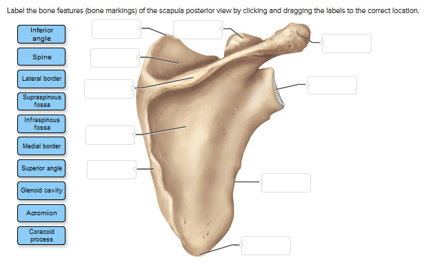
Name this specific part of the scapula. The scapula also known as the shoulder blade is a flat triangular bone located at the back of the trunk and resides over the posterior surface of ribs two to seven. Scapula Bone Introduction.
We created an anatomical atlas of the upper limb an interactive tool for studying the conventional anatomy of the shoulder arm forearm wrist and hand based on an axial magnetic resonance of the entire upper limb. There are six ossification centers of the elbow that appear and develop in a relatively reproducible fashion and are key to assessment of the pediatric elbow radiographTiming of their appearance varies in the literature but an approximation is given below. Name this specific part of the scapula.
Seventeen muscles attach to the scapula and it articulates with the clavicle to form the shoulder girdle or pectoral girdle which supports movements of the humerus. It floats on the rib cage and is attached to it only by muscles. The glenohumeral joint is between the head of the humerus and the _____ of the scapula.
The ulnar styloid can be seen posterior.

Scapula Bone Markings Anatomy Bones Scapula Medical Illustration

Left Scapula Posterior View Diagram Quizlet

Scapula Posterior View With Labels Appendicular Skeleton Visual Atlas Page 5 Human Anatomy And Physiology Anatomy And Physiology Shoulder Anatomy

The Pectoral Girdle Anatomy And Physiology I

Left Scapula Posterior View Labels 1 To 6 Indicate Areas Where Download Scientific Diagram

A P1lab Practical 2 Chapter 16 Flashcards Quizlet
Solved Label The Bone Features Bone Markings Of The Chegg Com

Scapula Posterior View Stock Photo Download Image Now Istock
0 comments
Post a Comment